


Antioxidants 2024, 13(5), 505; https://doi.org/10.3390/antiox13050505 (registering DOI) - 23 Apr 2024
Abstract
Astaxanthin (AST), functioning as an efficient antioxidant and pigment, is one of the most expensive additives in shrimp feeds. How to improve the uptake efficiency of dietary astaxanthin into farmed shrimp is of significance. The present study investigated the effects of lysophosphatidylcholine (LPC),
[...] Read more.
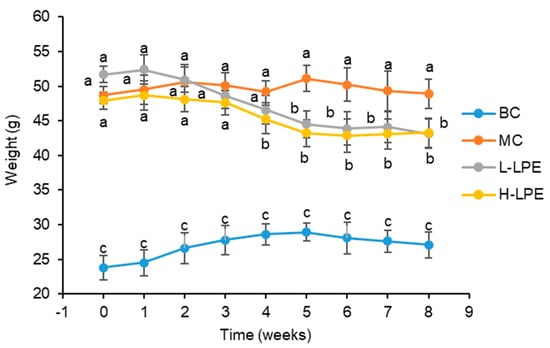
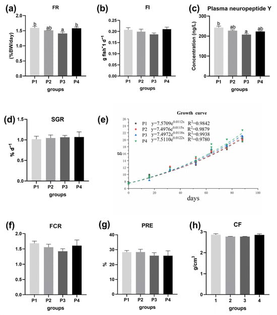
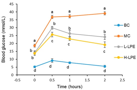
Astaxanthin (AST), functioning as an efficient antioxidant and pigment, is one of the most expensive additives in shrimp feeds. How to improve the uptake efficiency of dietary astaxanthin into farmed shrimp is of significance. The present study investigated the effects of lysophosphatidylcholine (LPC), an emulsifier, on dietary astaxanthin efficiency, growth performance, body color, body composition, as well as lipid metabolism of juvenile Pacific white shrimp (average initial body weight: 2.4 g). Three diets were prepared: control group, the AST group (supplemented with 0.02% AST), and the AST + LPC group (supplemented with 0.02% AST and 0.1% LPC). Each diet was fed to triplicate tanks, and each tank was stocked with 30 shrimp. The shrimp were fed four times daily for eight weeks. The AST supplementation improved the growth of white shrimp, while LPC further promoted the final weight of shrimp, but the whole-shrimp proximate composition and fatty acid composition were only slightly affected by AST and LPC. The LPC supplementation significantly increased the astaxanthin deposition in the muscle. The LPC supplementation significantly increased the shell yellowness of both raw and cooked shrimp compared to the AST group. Moreover, the dietary LPC increased the high-density lipoprotein-cholesterol content but decreased the low-density lipoprotein-cholesterol content in the serum, indicating the possible regulation of lipid and cholesterol transport. The addition of astaxanthin significantly up-regulated the expression of npc2 in the hepatopancreas compared to the control group, while the addition of LPC down-regulated the expression of mttp compared to the AST group. In conclusion, the LPC supplementation could facilitate the deposition of dietary astaxanthin into farmed shrimp and further enlarge the beneficial effects of dietary astaxanthin. LPC may also independently regulate shrimp body color and cholesterol transportation. This was the first investigation of the promoting effects of LPC on dietary astaxanthin efficiency.
Full article
(This article belongs to the Special Issue Natural Antioxidants and Aquatic Animal Health)


