


Biosensors 2024, 14(5), 213; https://doi.org/10.3390/bios14050213 - 24 Apr 2024
Abstract
The Wearable Robotic Limb (WRL) is a type of robotic arm worn on the human body, aiming to enhance the wearer’s operational capabilities. However, proposing additional methods to control and perceive the WRL when human limbs are heavily occupied with primary tasks presents
[...] Read more.
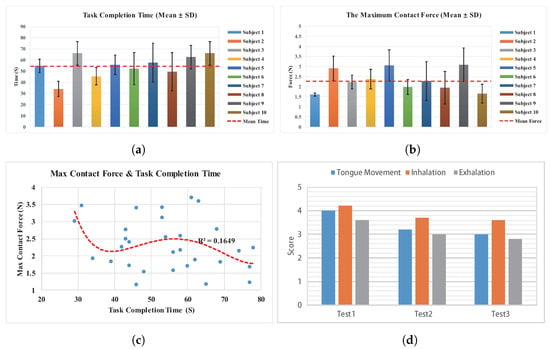
The Wearable Robotic Limb (WRL) is a type of robotic arm worn on the human body, aiming to enhance the wearer’s operational capabilities. However, proposing additional methods to control and perceive the WRL when human limbs are heavily occupied with primary tasks presents a challenge. Existing interactive methods, such as voice, gaze, and electromyography (EMG), have limitations in control precision and convenience. To address this, we have developed an interactive device that utilizes the mouth and tongue. This device is lightweight and compact, allowing wearers to achieve continuous motion and contact force control of the WRL. By using a tongue controller and mouth gas pressure sensor, wearers can control the WRL while also receiving sensitive contact feedback through changes in mouth pressure. To facilitate bidirectional interaction between the wearer and the WRL, we have devised an algorithm that divides WRL control into motion and force-position hybrid modes. In order to evaluate the performance of the device, we conducted an experiment with ten participants tasked with completing a pin-hole assembly task with the assistance of the WRL system. The results show that the device enables continuous control of the position and contact force of the WRL, with users perceiving feedback through mouth airflow resistance. However, the experiment also revealed some shortcomings of the device, including user fatigue and its impact on breathing. After experimental investigation, it was observed that fatigue levels can decrease with training. Experimental studies have revealed that fatigue levels can decrease with training. Furthermore, the limitations of the device have shown potential for improvement through structural enhancements. Overall, our mouth and tongue interactive device shows promising potential in controlling the WRL during tasks where human limbs are occupied.
Full article
(This article belongs to the Special Issue Devices and Wearable Devices toward Innovative Applications)
►
Show Figures

Figure 1


